
Professional Information
EYESIS Dry Eye Analyzer: A Guide
2024-10-19
Dry eye
The EYESIS dry eye analyzer (also known as the Ocular Surface Comprehensive Analyzer), as an emerging diagnostic tool, provides important support for the clinical diagnosis and assessment of dry eye. However, in practice, challenges remain, including non-standardized examination procedures, inappropriate selection of indications, and insufficient analysis of examination results. This article elaborates on the multiple non-invasive examination functions of the dry eye inspection and emphasizes the necessity of accurately interpreting examination findings and appropriately supporting clinical decision-making. In addition, a comparison between traditional examination methods and EYESIS dry eye analyzer is provided, and future directions of technological development are discussed。

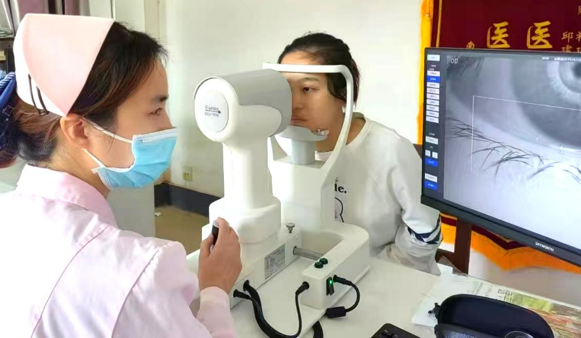
EYESIS dry eye analyzer, also known as the "Ocular Surface Comprehensive Analyzer," is a class of non-invasive ocular surface examination device with automated imaging and analysis capabilities. It offers multiple examination and analysis functions, including tear meniscus height measurement, non-invasive tear break-up time (NITBUT) assessment, lipid layer evaluation, and meibomian gland imaging.
The examination sequence follows the principle of performing less disruptive tests first, followed by more invasive/interfering tests. The recommended routine order is: tear meniscus height measurement → non-invasive tear break-up time assessment → lipid layer evaluation → meibomian gland imaging. If additional examination items are included, they may be inserted after the lipid layer assessment and before the meibomian gland examination.
For example, the default examination sequence of the EYESIS® Dry Eye Analyzer is as follows (the sequence can also be manually customized according to clinical needs)
1. Tear meniscus height measurement
2. Non-invasive tear break-up time (NITBUT)
3. Lipid layer assessment
4. Blink analysis
5. WTW (white-to-white) measurement
6. Redness analysis
7. Ocular surface staining (corneal staining)
8. Lid margin examination
9. Meibomian gland examination
10. OSDI
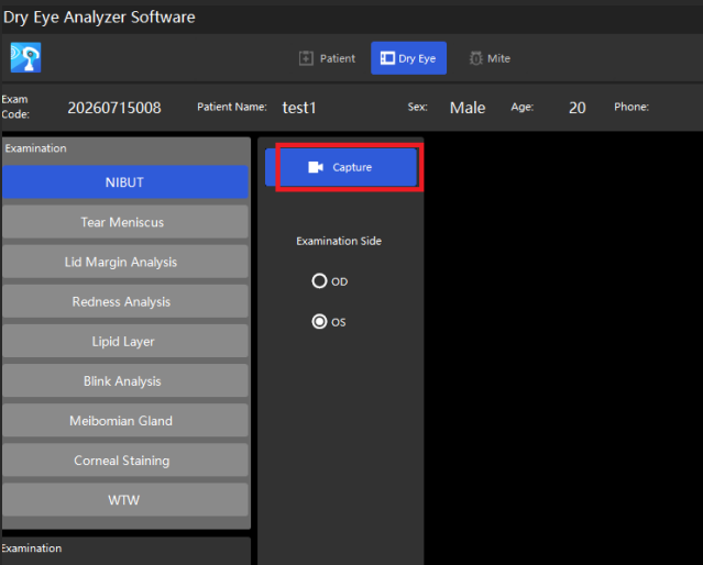
EYESIS Analyzer Item Sequence (Manually Adjustable)
1、TMH(Tear Meniscus Height)
(1)Inspection Principle:
TMH (Tear meniscus height) typically refers to the vertical height of the central lower tear meniscus at the coronal plane, below the pupillary center, and to some extent reflects the quantity of tear fluid. The Ocular Surface Comprehensive Analyzer first captures an image focused on the lower tear meniscus under white or near-infrared light, then manually or automatically delineates the upper and lower boundaries of the lower tear meniscus below the pupillary center. Based on these boundaries, the software calculates the TMH value.
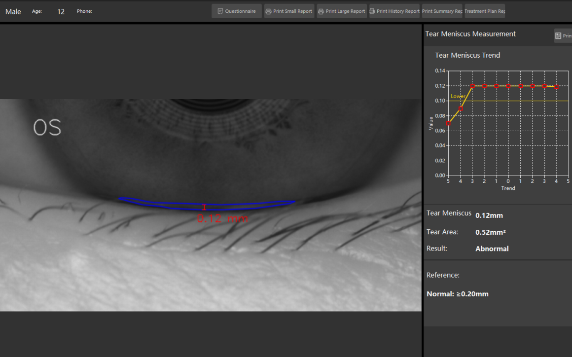
(2)Result Interpretation:
①Lower tear meniscus height > 0.2 mm: normal
②Lower tear meniscus height ≤ 0.2 mm: abnormal — one of the diagnostic indicators for dry eye
③Lower tear meniscus height ≤ 0.1 mm: moderately abnormal
④Unmeasurable: severely abnormal
(3)Precautions:
①Near-infrared light is recommended for TMH measurement, as white light stimulates tear secretion and may result in an artificially elevated TMH.
②Conjunctivochalasis can affect the identification of tear meniscus boundaries; loose bulbar conjunctiva stacked in the tear meniscus area may create a false elevation of TMH.
③ Irregular lid margins may cause distortion or discontinuity of the tear meniscus; this phenomenon is more prominent in patients with reduced tear secretion. Therefore, a smooth portion of the tear meniscus near the central region should be selected for measurement. ④Lacrimal drainage disorders, such as abnormalities of the lacrimal punctum, canaliculus, or nasolacrimal duct, can cause tear fluid accumulation and an elevated TMH.
⑤ It is recommended to avoid using eye drops, ophthalmic gels, or eye ointments for two hours before the examination.
(4)Comparison with Other Methods:
Traditional TMH measurement methods include slit-lamp microscopy estimation and fluorescein-stained slit-lamp microscopy estimation. Direct estimation using a slit-lamp microscope typically requires an eyepiece with a graticule to maximize accuracy. Since directly identifying the upper boundary of the lower tear meniscus is challenging, some studies add a small amount of fluorescein to the tear film to assist in visualizing the upper boundary. However, this invasive procedure may interfere with tear secretion and cause an artificially elevated TMH.
Unlike TMH measurement, the Schirmer test measures basal and reflex lacrimal gland secretion in an invasive manner. The Schirmer I test is performed without topical anesthetic. The basal Schirmer test, also known as the Jones test, uses topical anesthetic. The Schirmer II test, performed without topical anesthesia, additionally stimulates the nasal mucosa with a small cotton swab. The Schirmer test stimulates the ocular surface during the procedure, resulting in high variability and no correlation with TMH. Both TMH and the Schirmer I test are diagnostically meaningful for aqueous-deficient dry eye; they should complement rather than replace each other.
2、NIBUT(NIBUT)
(1)Principle of Examination:
The tear film is a thin film formed by tear fluid on the ocular surface. The tear film break‑up time refers to the time interval from blink completion to the appearance of the first break‑up spot in the tear film. It serves as a key indicator reflecting tear‑film stability and is also one of the diagnostic parameters for dry eye disease. Non‑invasive break‑up time (NIBUT) does not require fluorescein staining. Instead, patterns are projected onto the corneal area to observe the time of pattern distortion. At present, a Placido disc is commonly used for projection. The program automatically identifies the distorted position of the Placido‑disc reflection and records the distortion time.

(2)Result Analysis:
Tear‑film break‑up time is one of the important indicators for dry eye disease diagnosis. Taking NIBUT <10 s as the diagnostic threshold, the sensitivity ranges from 82 % to 84 % and the specificity ranges from 76 % to 94 %. NIBUT <5 s indicates moderate abnormality; NIBUT <2 s or absence of intact tear film indicates severe abnormality. NIBUT can be acquired by various devices. Some devices can obtain two parameters in a single examination: first NIBUT and average NIBUT. The first NIBUT refers to the time when the first break‑up spot appears during tear‑film disruption. The average NIBUT is the mean value of tear‑film break‑up time measured at multiple positions. Clinically, the first NIBUT is used as the diagnostic indicator for dry eye disease, and it is recommended to perform three measurements and take the average value.
(3)Precautions:
①Near‑infrared light is recommended for detection. White light makes it difficult for patients to maintain eye‑opening status.
②Accurate measurement results may not be obtained in patients with ptosis and/or corneal irregularity.
③The projection range of the Placido disc shall cover the entire cornea as far as possible.
④ It is recommended not to use eye drops, ophthalmic gels or ophthalmic ointments within two hours before examination.
(4)Comparison with Other Methods:
Fluorescein breakup time (FBUT) is the conventional method for measuring tear‑film stability. FBUT is an invasive examination; fluorescein can alter tear‑film conditions. There remains controversy regarding the magnitude of variability between FBUT and NIBUT. The correlation between FBUT and NIBUT is weak, and the two methods cannot substitute for each other. When NIBUT results differ substantially from clinical signs, combined FBUT examination is recommended for further evaluation.
3、Meibum quality
(1)Principle of Examination:
The lipid layer constitutes an essential component of the tear film. Analysis of the tear‑film lipid layer is based on the principle of thin‑film interference. Upon illumination with white light, the upper and lower interfaces of the lipid layer each reflect white light. The reflected light interferes with one another to produce multicolored fringes, namely the interference pattern. Tear‑film lipid‑layer analysis can be divided into qualitative analysis and quantitative analysis. The built‑in software of some devices can automatically calculate the lipid‑layer thickness over the underlying corneal surface by analyzing the colors of the interference pattern.
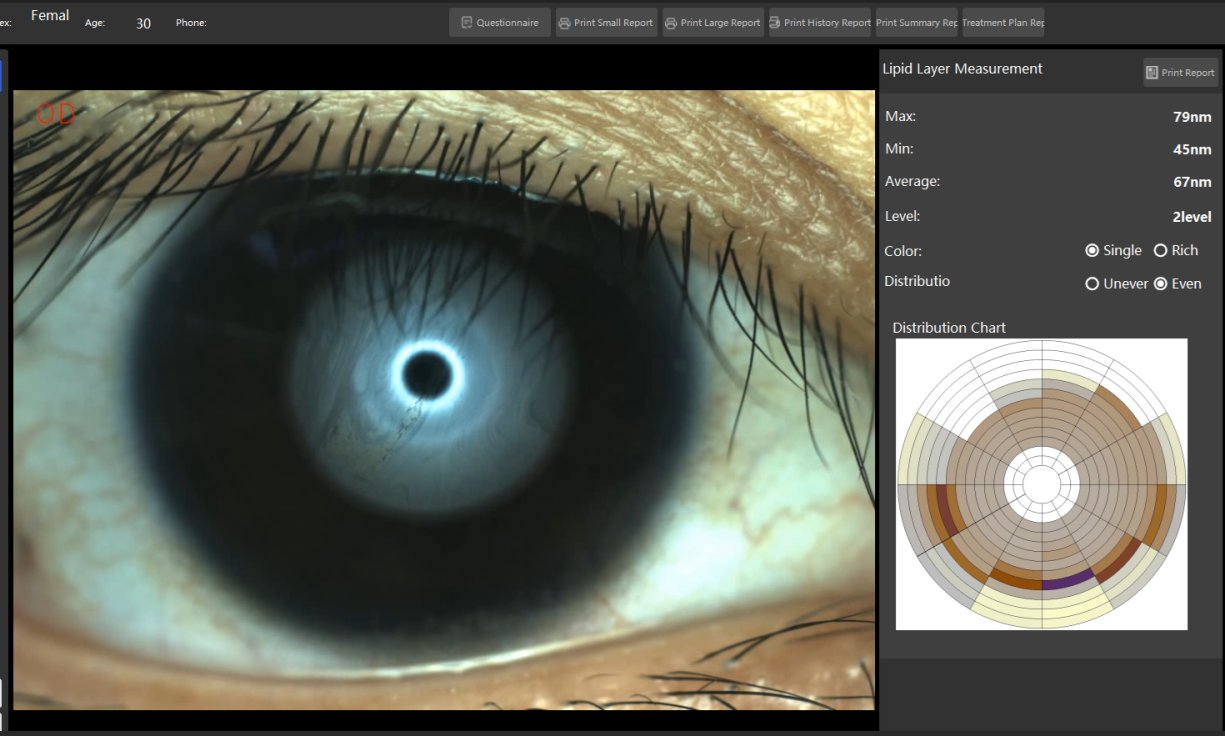
(2)Result Interpretation:
There are considerable differences between qualitative and quantitative analysis of the tear‑film lipid layer. Classification criteria for interference patterns vary widely among different interferometers, making a unified reference standard unavailable. Analysis can be performed from multiple dimensions including color, fluidity and morphology. A normal lipid layer presents uniform distribution with rich coloration. Quantitative parameters of the tear‑film lipid layer possess diagnostic value for meibomian gland dysfunction (MGD). According to published literature, when tear‑film lipid‑layer thickness ≤ 60 nm is adopted as the diagnostic criterion for MGD, the sensitivity is 47.9 % and the specificity is 90.2 %.
(3)Precautions:
① Patients should avoid oily cosmetics around the eyes before examination.
②It is recommended not to use eye drops, ophthalmic gels or ophthalmic ointments within two hours before examination.
③Do not rub or squeeze the eyes forcefully prior to testing.
4、Blink Analysis
(1)Principle of Examination:
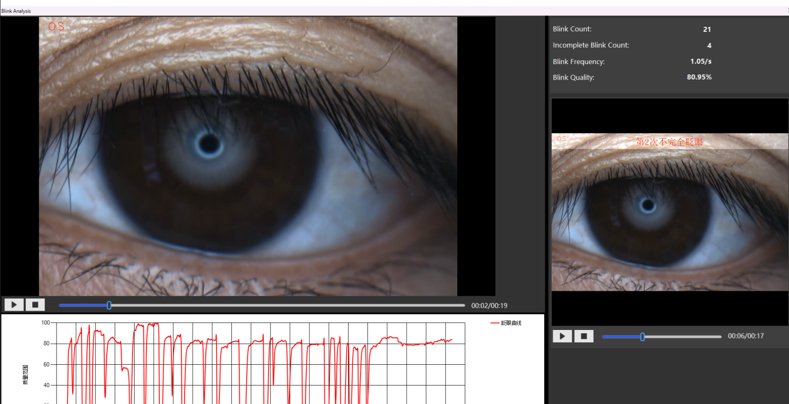
Blinking plays a vital role in maintaining a healthy ocular surface. Most blinks in healthy individuals throughout the day are spontaneous blinks. Blink examination mainly observes the frequency and completeness of patients’ spontaneous blinks. Based on whether the upper and lower eyelids make contact during a single blink, blinks can be categorized as complete blinks and incomplete blinks.
(2)Result Interpretation:
The device adopts partial blink (PB) ratio as the blink‑related parameter. PB is presented as a fraction in the report. The denominator stands for the total number of blinks within a unit time (20 s), and the numerator represents the number of incomplete blinks within the same period. Therefore, both the total blink count per unit time and PB should be considered. The spontaneous blink frequency in normal adults is approximately 10‑20 blinks per minute, equivalent to 3‑7 blinks /20 s. Abnormal blinking includes abnormalities in blink frequency and blink completeness. Abnormal blinking frequently occurs during reading, working and video‑display‑terminal use, and becomes aggravated with increased reading or work‑related load. In addition, abnormal blinking may result from neurological disorders such as Parkinson’s syndrome and facial nerve dysfunction. Ocular diseases including thyroid‑associated ophthalmopathy, Meige syndrome, conjunctivitis and refractive errors can also induce abnormal blinking. Partial corneal nerve transection during cataract surgery and refractive surgery may reduce postoperative blink frequency. Eyelid surgeries such as double‑eyelid blepharoplasty and ptosis correction may lead to lagophthalmos and consequently incomplete blinks.
(3)Precautions:
(1) Explain the purpose and procedure of the examination to the patient to ensure natural and relaxed blinking during testing. (2) Collect information on the subject’s occupation, living habits and daily video‑display‑terminal usage duration before examination. (3) Ambient lighting may influence measurement results. It is recommended to perform the test under bright indoor illumination.
(4)Comparison with Other Methods:
Videography is commonly used for blink assessment. Observers perform subjective analysis of blink movements by watching slow‑motion video playback. Variations in camera frame rate and inter‑observer differences can both affect test outcomes.
5、Meibomian Gland Analysis
(1)Principle of Examination:
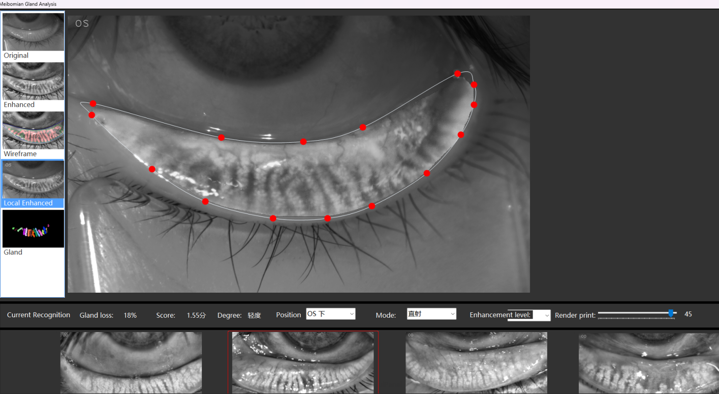
Meibomian glands are sebaceous glands located within the eyelid. The glands are arranged in parallel and run perpendicular to the eyelid margin. In normal individuals, the upper eyelid contains 20‑40 meibomian glands, with an average of 31; the lower eyelid contains 20‑30 meibomian glands, with an average of 26. Meibomian glands can also be visualized under a slit‑lamp microscope. However, the ocular surface comprehensive analyzer adopts multiple imaging modes including near‑infrared illumination and adaptive transillumination to achieve clearer visualization of meibomian glands. Physicians can observe finer anatomical details such as meibomian gland acini, central ducts and secretory ducts.
(2)Result Interpretation:
Meibomian gland loss grading can be adopted to evaluate meibomian gland morphology. Normal status is defined as no meibomian gland loss; mild loss corresponds to a gland‑loss ratio less than 1/3; moderate loss is defined by a loss ratio ranging from 1/3 to 2/3; severe loss refers to a gland‑loss ratio greater than 2/3. The degree of meibomian gland loss correlates with patients’ subjective symptoms, tear‑film break‑up time and meibomian gland function. In addition, other assessment approaches for meibomian gland morphology include meibomian gland distortion scoring and evaluation of gland thickness, yet these methods have not been widely applied. Artificial‑intelligence‑based methods have also been utilized for quantitative and objective analysis of meibomian gland morphology.
(3)Precautions:
①Evert the eyelid fully to expose as much meibomian gland tissue as possible.
② Keep the everted eyelid flat.
③After eversion, the palpebral conjunctival surface shall be perpendicular to the light source to acquire clear images.
④Eyelid eversion may be difficult in patients after eyelid surgery.
⑤For patients with prior eyelid surgery, meibomian gland loss may be present at the surgical site.
6、Redness
(1)Principle of Examination:
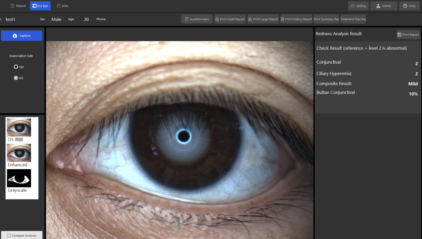
Conjunctival redness grade is one of the indicators for assessing ocular‑surface inflammation. After the ocular surface comprehensive analyzer captures images of the bulbar conjunctiva and/or limbus, its built‑in software can automatically detect the visible nasal and temporal bulbar conjunctiva. The degree of redness is estimated by quantifying the ratio of the vessel‑covered area to the total measured area.
(2)Result Interpretation:
The ocular surface comprehensive analyzer outputs conjunctival redness scores for the nasal and temporal bulbar conjunctiva. Some devices also provide an overall redness score calculated as a weighted average based on the nasal‑side and temporal‑side redness grades and the analyzed regions. At present, there is no unified grading standard for conjunctival redness.
(3)Precautions:
Communicate fully with the patient before measurement. Instruct the patient to keep the eyes open as much as possible during measurement to expose the bulbar conjunctiva.
(4)Comparison with Other Methods:
Subjective redness scoring relies mainly on observations under a slit‑lamp microscope. Test results can be influenced by differences in observers’ experience.
7、Ocular Surface and Eyelid Margins

At present, the ocular surface comprehensive analyzer can directly assess the status of the aqueous and lipid layers of the tear film via tear meniscus height measurement and tear‑film lipid‑layer examination, and evaluate the tear film as a whole through non‑invasive break‑up time (NIBUT) testing.
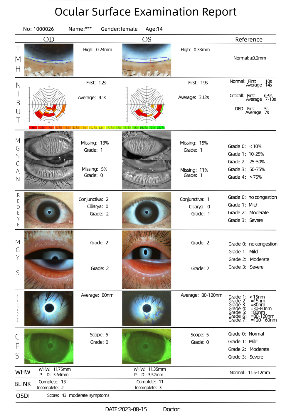
EYESIS Dry Eye Analyzer Report
The advent of the EYESIS dry eye analyzer has, to a certain extent, addressed the drawbacks such as invasiveness and subjectivity that exist in conventional dry‑eye examinations. Nevertheless, limitations still remain. In clinical practice, clinicians should correctly interpret the various parameters obtained from the ocular surface comprehensive analyzer, understand the clinical significance of each parameter, and combine these findings with traditional dry‑eye examination modalities to achieve better assessment of the patient’s ocular‑surface condition. In addition, more intelligent ocular surface comprehensive analyzers and the establishment of diagnostic prediction models can be expected in the future.